 Written and reviewed by Dr. Vaishnavi Karmawat, B.V.Sc. & A.H., Registered Veterinary Practitioner
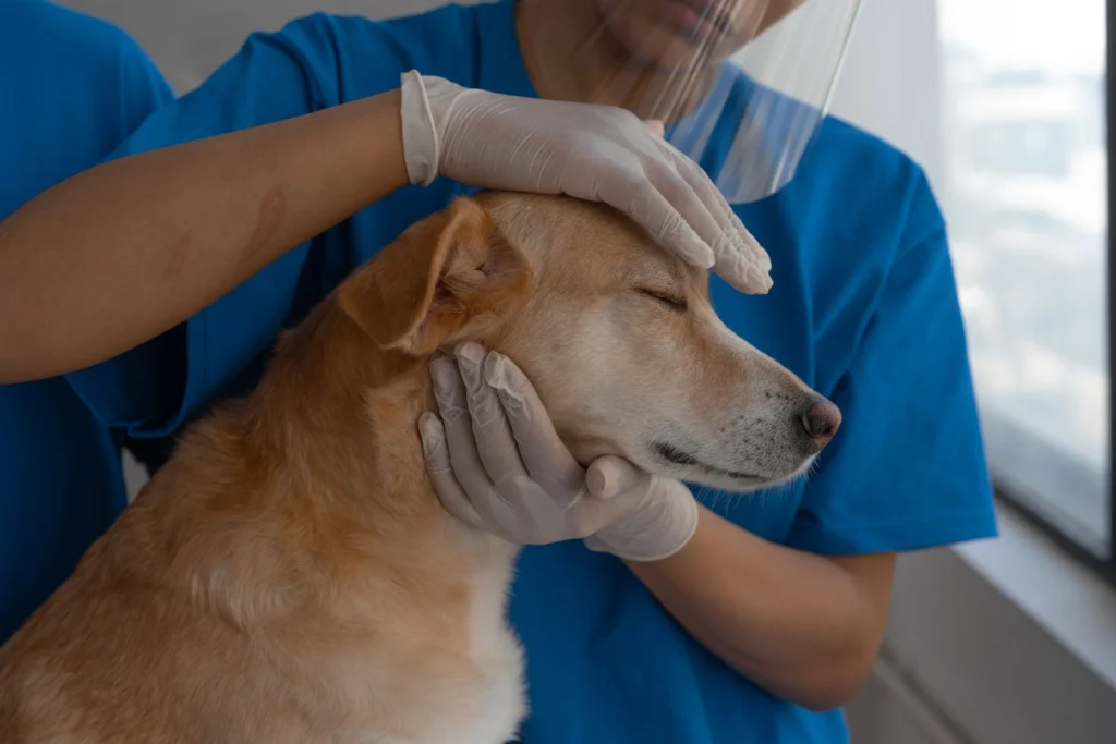
Short answer: most lumps on dogs are harmless: fatty lumps (lipomas), cysts, warts and skin tags. But some cancers, especially mast cell tumours, can look and feel exactly like a harmless lump. No one, including a vet, can tell what a lump is just by looking or feeling. A quick, inexpensive needle test (fine-needle aspirate) in the clinic often gives the answer the same day. Any new lump that is bigger than a pea (about 1 cm), has been there for a month, or is changing should be checked.
In this article
Key takeaways
- Follow “see something, do something”: measure, photograph and get any lump over about 1 cm, or present for over a month, sampled by a vet.
- Fine-needle aspiration (FNA) and cytology is quick, cheap and usually needs no sedation. A biopsy gives a definite diagnosis and grade when FNA is unclear.
- Mast cell tumours are the most common skin cancer in dogs. They can swell and shrink, look like a bug bite or a fatty lump, and release histamine when handled.
- Early removal of a cancerous lump with adequate margins is often curative, and smaller lumps need smaller, cheaper surgery.
Common benign (non-cancerous) lumps
| Lump | What it feels and looks like | Typical dogs | What to do |
|---|---|---|---|
| Lipoma (fatty tumour) | Soft, squishy, movable, under the skin; chest, belly, armpits | Middle-aged to older, overweight dogs; Labradors | Confirm with FNA; monitor size. Remove if large, fast-growing, or affecting movement. Infiltrative lipomas grow into muscle |
| Sebaceous cyst / adenoma | Small, firm, wart-like or cauliflower-like; may ooze a white, cheesy material | Older dogs; Poodles, Cocker Spaniels, Shih Tzus | Leave alone or remove if irritated or bleeding; do not squeeze |
| Viral papilloma (wart) | Pink-grey cauliflower growths in or around the mouth, eyelids, paws | Young dogs, dogs with weak immunity | Usually regress in 1–5 months; remove if bleeding or blocking eating |
| Histiocytoma (“button tumour”) | Round, red, hairless, raised button; head, ears, legs; grows fast | Dogs under 3 years | Usually regresses within 3 months; confirm with FNA (can mimic mast cell tumour) |
| Skin tag | Soft, fleshy, stalk-like | Older dogs | Harmless unless caught or bleeding |
| Abscess | Warm, painful, soft swelling that may burst with pus | After bites, thorns, injections | Vet lancing, flushing, antibiotics |
| Haematoma, seroma | Soft fluid-filled swelling after trauma or surgery | Any | Usually resolves; vet check |
| Engorged tick | Grey-brown, bean-like, with legs near the skin | Any; very common in India | Remove carefully with a tick tool, and start tick control. See tick fever |
Cancerous (malignant) lumps to know about
| Tumour | Clues | Notes |
|---|---|---|
| Mast cell tumour (MCT) | Any appearance: can be soft, firm, red, itchy, ulcerated; may swell and shrink, especially after being touched (histamine release, called Darier’s sign) | Most common skin cancer in dogs; Boxers, Pugs, Labradors, Golden Retrievers, Boston Terriers prone; graded low or high by biopsy; can spread to lymph nodes, spleen and liver |
| Soft tissue sarcoma | Firm, fixed, under the skin; slow-growing; limbs and trunk | Locally invasive “finger-like” roots; needs wide surgical margins |
| Melanoma | Dark pigmented lump; in the mouth or toes/nail beds often malignant; on haired skin often benign | Oral and digit melanomas spread early |
| Squamous cell carcinoma | Crusty, ulcerated, non-healing sore; pale skin areas, nose, belly, nail beds | Linked to sun exposure in pale-skinned dogs |
| Mammary tumours | Lumps along the teats in females | About half are malignant in dogs; very common in unspayed females; spaying before the first or second heat greatly reduces the risk |
| Lymphoma | Several firm, painless swellings under the jaw, in front of the shoulders and behind the knees (lymph nodes) | Systemic cancer; responds well to chemotherapy initially |
| Transmissible venereal tumour (TVT) | Cauliflower-like, bleeding masses on the genitals, sometimes nose or mouth | Common in Indian street and breeding dogs; spread by mating, licking and sniffing; responds very well to vincristine chemotherapy |
How vets test a lump

- Measure and map: size (in cm), location, firmness, attachment, photos. Your vet will record it on a body map.
- Fine-needle aspirate (FNA) and cytology: a thin needle collects cells, which are stained and examined in the clinic or sent to a pathologist. It distinguishes fat, cysts, inflammation, mast cells and many tumours. It usually takes minutes, with no sedation.
- Biopsy: a small piece (punch or wedge biopsy) or the whole lump (excisional biopsy) is examined by histopathology for a definite diagnosis, tumour grade and, after removal, surgical margins.
- Staging (if cancer): lymph node aspirates, blood tests, chest X-rays, abdominal ultrasound to check for spread.
- Before surgery for a mast cell tumour, vets give antihistamines and stomach protectants, because handling the tumour releases histamine.
Treatment
- Benign lumps: often just monitored, or removed if large, ulcerated, irritating or in an awkward place.
- Surgical removal is the main treatment for most skin cancers. Planned margins matter: for mast cell tumours and sarcomas, vets remove a rim of normal tissue (often 2–3 cm laterally and one tissue layer deep).
- The histopathology report tells whether margins are “clean”. Incomplete margins may need a second surgery or radiotherapy.
- Chemotherapy for lymphoma, TVT, high-grade mast cell tumours, or tumours that have spread. Dogs tolerate chemotherapy much better than people, and most have few side effects.
- Targeted drugs: toceranib (Palladia) for some mast cell tumours; electrochemotherapy and radiotherapy in referral centres.
Checking your dog at home
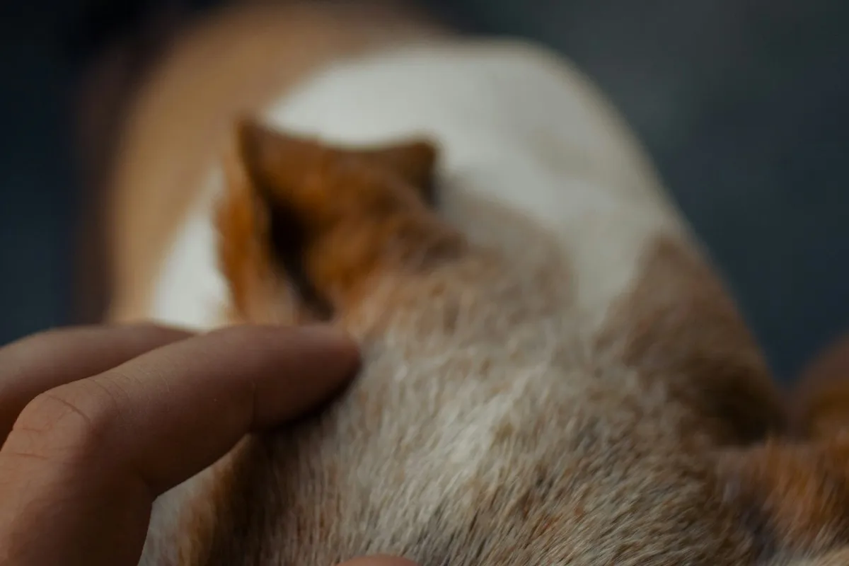
- Once a month, run your hands over your dog’s whole body: head, neck, chest, belly, legs, between the toes, tail, and along the teats.
- Look inside the mouth and at the gums.
- Feel the lymph nodes under the jaw, in front of the shoulders and behind the knees.
- Record new lumps: date, location and size. Measure with a ruler, and photograph with a coin for scale.
- Keep your dog at a healthy weight. Fat makes small lumps harder to feel.
Lumps in cats are different
Cats get fewer lumps than dogs, but a higher proportion of them are cancerous, so every new lump on a cat deserves a vet check.
| Lump | Clues | What to do |
|---|---|---|
| Abscess from a bite | Painful, warm swelling that appears within days of a fight, often with fever; may burst and drain pus | Vet visit for draining and antibiotics; test for FIV and FeLV later |
| Mammary (breast) tumour | Firm lumps along the belly near the nipples, usually in older unspayed females | Up to about 85 to 90 percent are malignant in cats. Early surgery is essential. Spaying before 6 months greatly reduces the risk |
| Injection-site sarcoma | Firm lump where a vaccine or injection was given, still present 3 months later, larger than 2 cm, or growing a month after the injection | Biopsy before surgery; wide removal is needed |
| Mast cell tumour | Small, firm, hairless bumps, often on the head and neck | Usually benign in the skin of cats, but needs testing |
| Squamous cell carcinoma | Crusty, non-healing sores on the ear tips, nose or eyelids of white cats exposed to strong sun | Early surgery or other treatment; keep white cats out of midday sun |
| Basal cell tumour | Single firm, often dark lump in older cats | Usually benign; removal is curative |
Frequently asked questions
My dog’s lump is soft and moves around. Is it just fat?
Often yes, but mast cell tumours can feel identical to a lipoma. Only cytology can confirm it. Have every new lump aspirated at least once.
Will a needle test spread the cancer?
The risk is extremely small, and the information gained far outweighs it. It is a standard, safe procedure.
Can lumps be prevented?
Not all of them. Spaying early prevents most mammary tumours; sun protection helps pale dogs; tick control prevents tick lumps and infections; keeping dogs lean reduces lipomas.
My old dog has many lumps. Do they all need surgery?
No. Your vet will sample new or changing lumps, and many benign ones can simply be monitored. Decisions are based on the diagnosis, growth, location and your dog’s overall health.
Is TVT contagious to other dogs?
Yes, it spreads between dogs through mating and close contact with the tumour. It is not contagious to people, and it usually responds very well to chemotherapy.
When to call a vet
See a vet for any lump larger than a pea, present for a month, growing, changing shape or colour, ulcerating, bleeding or itchy, and for any lump in the mouth, on the toes, along the mammary chain, or multiple swollen lymph nodes. Go promptly for a hot, painful swelling.
Related Articles
- Is My Dog or Cat Dehydrated? 1-Minute Gum and Skin Tent Check, ORS and When To See a Vet
- Bloody Diarrhoea in Dogs (Raspberry Jam Stool): Colitis vs Hookworms vs Parvo vs AHDS in Indian Dogs
- Dog Pooping Blood in India: Red Blood (Hematochezia) vs Black Tarry Stool (Melena), Causes and When To See a Vet
- Dog Vomiting: Colour Guide, Causes, Vet Treatment and When To Worry
References
- Withrow SJ, Vail DM, Thamm DH. Withrow and MacEwen’s Small Animal Clinical Oncology, 6th ed. Elsevier.
- Kiupel M, et al. Proposal of a 2-tier histologic grading system for canine cutaneous mast cell tumors. Vet Pathol 2011;48:147–155.
- Veterinary Society of Surgical Oncology (VSSO): “See Something, Do Something: Why Wait? Aspirate.” campaign.
- Ganguly B, et al. Canine transmissible venereal tumour: a review. Vet Comp Oncol 2016.
- Merck Veterinary Manual: Tumors of the skin and soft tissues in dogs.
- Hayes HM, Milne KL, Mandell CP. Epidemiological features of feline mammary carcinomas. Veterinary Record. 1981;108(22):476-479.
More on Dog Diseases and Infections

Metronidazole (Metrogyl, Flagyl) for Dogs and Cats: When Vets Use It for Loose Motion and Giardia, and Why Overdose Causes Wobbling
Metronidazole for dogs and cats: uses for Giardia and gut infections, when it is not needed, giving the bitter pill, and neurotoxicity…

Congestive Heart Failure in Dogs: Night Coughing, Sleeping Breath Count, Stages and Heart Care for Senior Dogs
Heart failure in dogs: mitral valve disease vs DCM, stages, murmurs, cough and fast breathing, medicines, and counting sleeping breaths at home.